Diagram Of Chest Area / Knee Muscles Anatomy, Function & Diagram | Body Maps : Thoracic cavity, also called chest cavity, the second largest hollow space of the body.it is enclosed by the ribs, the vertebral column, and the sternum, or breastbone, and is separated from the abdominal cavity (the body's largest hollow space) by a muscular and membranous partition, the diaphragm.it contains the lungs, the middle and lower airways—the tracheobronchial tree—the heart.
Diagram Of Chest Area / Knee Muscles Anatomy, Function & Diagram | Body Maps : Thoracic cavity, also called chest cavity, the second largest hollow space of the body.it is enclosed by the ribs, the vertebral column, and the sternum, or breastbone, and is separated from the abdominal cavity (the body's largest hollow space) by a muscular and membranous partition, the diaphragm.it contains the lungs, the middle and lower airways—the tracheobronchial tree—the heart.. Inside, heart is hollow and divided into 4 chambers: It starts from the pharynx and extends to the upper end of the esophagus. Nerves of the chest and upper back. Any diaphragm pain can, therefore, be very alarming. A spasm may feel like a twitch or flutter and can occur with or without pain.
The heart is enclosed in the pericardium which is a double layer. The chest and the abdomen are separated by a large muscle called the diaphragm, the major breathing muscle. Possible causes of pain include trauma, musculoskeletal. Of the two chest muscles, the pectoralis major (a.k.a. 9 / 10 ( 1 vote ) location of chest pain during angina or heart attack diagram.
The Bodywork Perspective - the Chest and Sternum - The ... from phoenixcentre.com The heart is enclosed in the pericardium which is a double layer. Chest area diagram chest pain area diagram lymph nodes in chest area diagram. Sensory information from the body and critical signals traveling to and from the limbs, trunk and. The muscles of the hiatus form a sphincter around the food pipe that. A diaphragm spasm is an involuntary contraction of the muscle that divides the upper abdomen and chest. This pericardium is attached to the diaphragm, spinal column and other parts via strong ligaments. Other major causes of pain in right chest area are: The ribs and sternum make up what is called the 'ribcage.' the ribcage protects the lungs, blood vessels, and heart.
The throat is one of the most complex parts of the human body.
For many, the chest is made up of a single rigid bone called the sternum.however, this is not true.other than the sternum, there are other bones in the chest region, such as the ribs and even the spine at the back. System respiratory respiratory organs of human body digestive and respiratory system medical chest internal structure of human body medicine body lungs biology intestines stomach anatomy torso human internal. The diaphragm, a sheet of muscle in the middle chest area, is essential for breathing. Based on anatomy, throat can be divided into 3 parts namely, the upper part, the middle part and the lower part called as nasopharynx, oropharynx and laryngopharynx respectively. The circulatory system does most of its. This aiming spot will place the arrow at the top of the heart and center of both lungs. The throat is responsible for performing a large number of functions, namely the swallowing, speaking and breathing. The sternum, or breastbone, is a flat bone at the front center of the chest. Possible causes of pain include trauma, musculoskeletal. The throat is one of the most complex parts of the human body. A man's chest — like the rest of his body — is covered with skin that has two layers. It starts from the pharynx and extends to the upper end of the esophagus. 1/3 to 1/2 up the chest cavity.
This thoracic and pulmonary anatomy tool is especially designed for students of anatomy (medical and paramedical studies). Remember, an arrow will penetrate through the ribs. It lies between the right and left lungs, in the middle of the chest and slightly towards the left of the breastbone. In this image, you will find an upper chest, substernal radiating to neck and jaw, substernal raiding down left arm, substernal radiating down left arm, epigastric radiating to neck, jaw, and arms, neck and jaw, left shoulder and down both arms, intrascapular in it. Chest area diagram chest pain area diagram lymph nodes in chest area diagram.
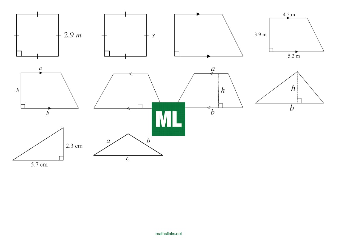
Perimeter and Area - diagram library - MathsFaculty from mathslinks.imgix.net Possible causes of pain include trauma, musculoskeletal. The pec major) is the one that commands the most real estate. Find the perfect male chest anatomy stock photo. Human chest bone structure parts of the chest bones. Any diaphragm pain can, therefore, be very alarming. The throat is responsible for performing a large number of functions, namely the swallowing, speaking and breathing. No need to register, buy now! It lies between the right and left lungs, in the middle of the chest and slightly towards the left of the breastbone.
Other major causes of pain in right chest area are:
Of the two chest muscles, the pectoralis major (a.k.a. It lies between the right and left lungs, in the middle of the chest and slightly towards the left of the breastbone. Possible causes of pain include trauma, musculoskeletal. If the front legs are spread apart, follow up the of the legs 1/3 to 1/2 up the chest. The chest is the area of origin for many of the body's systems as it houses organs such as the heart, esophagus, trachea, lungs, and thoracic diaphragm. Anatomy of the chest and the lungs: Human chest bone structure parts of the chest bones. The circulatory system does most of its. The muscles of the hiatus form a sphincter around the food pipe that. A diaphragm spasm is an involuntary contraction of the muscle that divides the upper abdomen and chest. For many, the chest is made up of a single rigid bone called the sternum.however, this is not true.other than the sternum, there are other bones in the chest region, such as the ribs and even the spine at the back. The sternum, or breastbone, is a flat bone at the front center of the chest. Find the perfect male chest anatomy stock photo.
Anatomy of the chest and shoulder, anatomy of the chest organs, anatomy of the chest wall, anatomy of the chest wall and pleura, anatomy of upper chest area, human. Immediately following the pharynx are the larynx, epiglottis, larynx and the esophagus. For many, the chest is made up of a single rigid bone called the sternum.however, this is not true.other than the sternum, there are other bones in the chest region, such as the ribs and even the spine at the back. Human chest bone structure parts of the chest bones. The sternum, or breastbone, is a flat bone at the front center of the chest.
Glossary - ADAPT from www.adaptacademy.com A diaphragm spasm is an involuntary contraction of the muscle that divides the upper abdomen and chest. The chest is the area of origin for many of the body's systems as it houses organs such as the heart, esophagus, trachea, lungs, and thoracic diaphragm. Immediately following the pharynx are the larynx, epiglottis, larynx and the esophagus. Profile view of female chest area. 1/3 to 1/2 up the chest cavity. It lies between the right and left lungs, in the middle of the chest and slightly towards the left of the breastbone. Anatomy of the chest and the lungs: Possible causes of pain include trauma, musculoskeletal.
Both methods locate the aiming spot to put the arrow in the center of the vital organ area.
It starts from the pharynx and extends to the upper end of the esophagus. System respiratory respiratory organs of human body digestive and respiratory system medical chest internal structure of human body medicine body lungs biology intestines stomach anatomy torso human internal. Your pectoralis major and pectoralis minor muscles make up most of the muscle mass in your chest. Profile view of female chest area. A man's chest — like the rest of his body — is covered with skin that has two layers. If the front legs are spread apart, follow up the of the legs 1/3 to 1/2 up the chest. Nerves of the chest and upper back. The sternum is located near the heart, so many people. The nervous system of the thorax is a vital part of the nervous system as a whole, as it includes the spinal cord, peripheral nerves, and autonomic ganglia that communicate with and control many vital organs. No need to register, buy now! Immediately following the pharynx are the larynx, epiglottis, larynx and the esophagus. The circulatory system does most of its. Both methods locate the aiming spot to put the arrow in the center of the vital organ area.


Posting Komentar
0 Komentar